Calcified left adrenal hematoma
Home » » Calcified left adrenal hematomaYour Calcified left adrenal hematoma images are available in this site. Calcified left adrenal hematoma are a topic that is being searched for and liked by netizens now. You can Find and Download the Calcified left adrenal hematoma files here. Get all free photos and vectors.
If you’re searching for calcified left adrenal hematoma images information connected with to the calcified left adrenal hematoma keyword, you have pay a visit to the right blog. Our site always gives you suggestions for refferencing the highest quality video and image content, please kindly surf and locate more enlightening video articles and graphics that fit your interests.
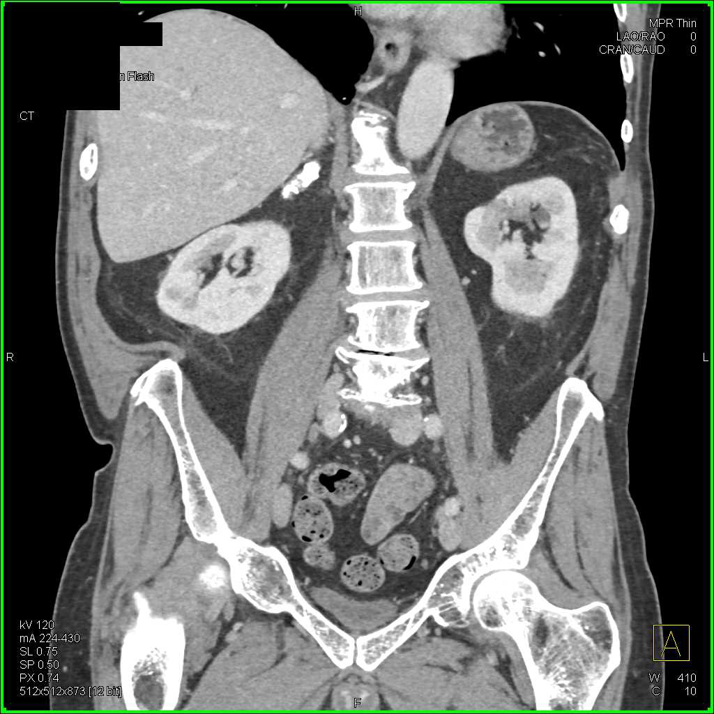
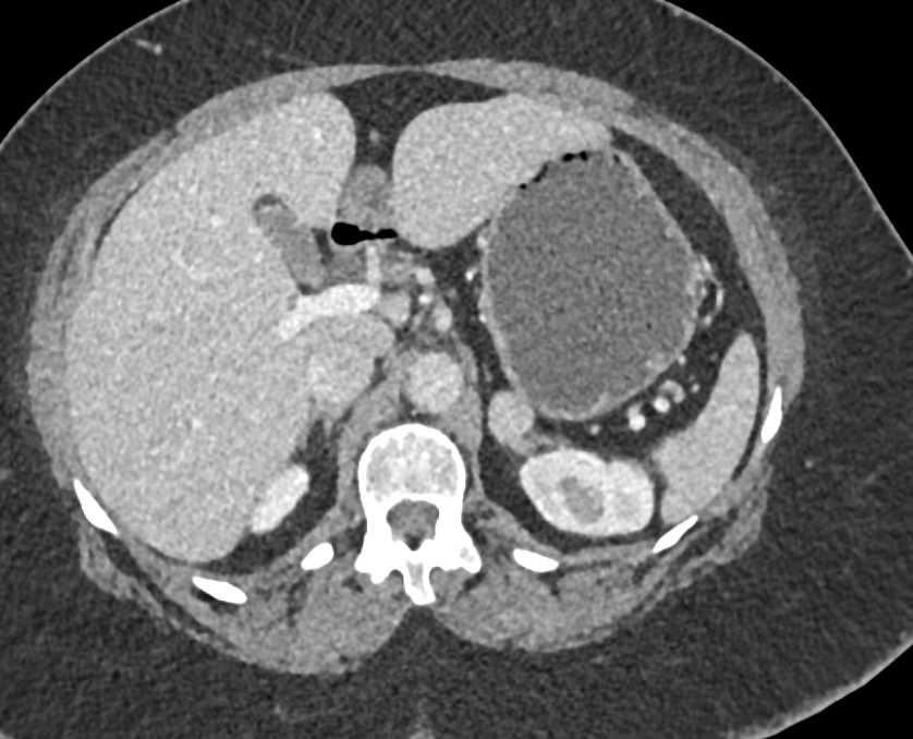
Calcified Left Adrenal Hematoma. Complication of adrenal gland injury includes adrenal vein rupture with haemorrhage both into the adrenal gland and in the retroperitoneum. B, unenhanced axial ct image shows calcified hematoma (asterisk) within left adrenal gland. A calcified hematoma is a nonhereditary form of heterotopic or myositis ossification (also known as osteogenesis, a form of bone remodelling) is a type of physical change of deep bruises. On the other hand, adenomas rarely calcify.
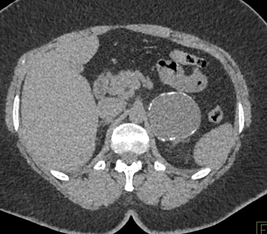 Partially calcified Old Hematoma of the Left Adrenal From ctisus.com
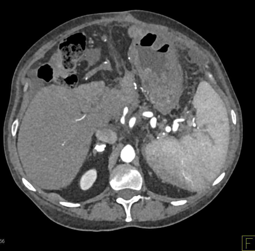
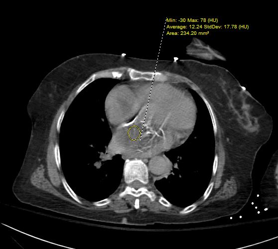
The etiologies for this unusual disorder are. Distension of the cranial sutures at all with meningiosis leucemica 2: It is characterized by a persistent hematoma that increases in size for more than one. This case has implications for the investigation of children with adrenal. Abdominal computer tomography (ct) showed 7 × 9 × 6 cm left adrenal mass with calcifications and central and peripheral enhancement with iv contrast, highly suspicious for primary adrenal carcinoma or metastatic disease (fig. The image shows a 7,6 cm left adrenal lesion with density < 0 hu, but with hyperdense strands.
Some adrenal haematomas may calcify after one year.
Adrenal hemorrhage is an uncommon disorder characterized by bleeding into the suprarenal glands. 5 the majority of adrenal gland. We report a case of adrenal calcification without a noncalcified mass in a child who subsequently presented with neuroblastoma elsewhere. Months later, she started to have abdominal pain in left upper quadrant (luq) radiating to her back. Complication of adrenal gland injury includes adrenal vein rupture with haemorrhage both into the adrenal gland and in the retroperitoneum. Video chat with a u.s.
 Source: ctisus.com
Adrenal metastases may have a similar appearance to the primary neoplasm. Lung abscess due to staphylococci 3: Adrenal calcification after adrenal hemorrhage other cases by these authors: Several months after the acute event, ct scanning of the adrenals shows progressive atrophy, with the variable appearance of calcifications. Concretions these are discrete precipitates in a vessel or organ.
 Source: ctisus.com
Adrenal trauma should not be considered an incidental finding because it can lead to severe bleeding, which may require a blood transfusion. [4] other methods are adopted as the patients grow because the imaging of adrenal gland would become more difficult with ultrasound. Bleeding often continues until the adrenal gland expands beyond the adreniform shape and forms a round or oval hematoma in the gland. The hematoma may be unilateral or bilateral, and the clinical presentation can range from nonspecific abdominal pain to catastrophic cardiovascular collapse. Distension of the cranial sutures at all with meningiosis leucemica 2:
 Source: ctisus.com
It is characterized by a persistent hematoma that increases in size for more than one. Adrenal calcification after adrenal hemorrhage other cases by these authors: Hematomas usually decrease in size and may spontaneously disappear. Spontaneous regression is followed by a classic rim of calcification leaving a normal sized calcified gland. The hematoma may be unilateral or bilateral, and the clinical presentation can range from nonspecific abdominal pain to catastrophic cardiovascular collapse.
 Source: ctisus.com
Video chat with a u.s. Lung abscess due to staphylococci 3: Hematomas usually decrease in size and may spontaneously disappear. B, unenhanced axial ct image shows calcified hematoma (asterisk) within left adrenal gland. The neonatal appearance falls into three main categories (5,6):
 Source: ctisus.com
Lung abscess due to staphylococci 3: It mostly takes place when people sustain deep bruises along with bleeding because of any forceful, blunt trauma such as getting kicked or having a car accident. Her biochemical workup showed no evidence of. Adrenal calcification after adrenal hemorrhage other cases by these authors: Adrenal trauma should not be considered an incidental finding because it can lead to severe bleeding, which may require a blood transfusion.
 Source: ctisus.com
Video chat with a u.s. When the adrenal gland is calcified but no mass is found, the calcification is usually assumed to be due to prior adrenal hemorrhage. Concretions these are discrete precipitates in a vessel or organ. The imaging workup of suspected neonatal adrenal hemorrhage typically begins with ultrasound. Abdominal computer tomography (ct) showed 7 × 9 × 6 cm left adrenal mass with calcifications and central and peripheral enhancement with iv contrast, highly suspicious for primary adrenal carcinoma or metastatic disease (fig.
 Source: heart.thecommonvein.net
Approximately 70% of adrenal hemorrhages occur on the right, a phenomenon thought to result from compression of the gland located between the liver and the kidney. Complication of adrenal gland injury includes adrenal vein rupture with haemorrhage both into the adrenal gland and in the retroperitoneum. They are sharp in outline but the density and shape vary but. Adrenal hemorrhage is an uncommon disorder characterized by bleeding into the suprarenal glands. Atypical adenoma with hyperdense strands as a result of internal bleeding.
 Source: ctisus.com
Calcification in adenomas occurs after. The neonatal appearance falls into three main categories (5,6): Adrenal hematoma is a rare diagnosis for large adrenal masses. Abdominal computer tomography (ct) showed 7 × 9 × 6 cm left adrenal mass with calcifications and central and peripheral enhancement with iv contrast, highly suspicious for primary adrenal carcinoma or metastatic disease (fig. Distension of the cranial sutures at all with meningiosis leucemica 2:
 Source: ctisus.com
Video chat with a u.s. Bleeding often continues until the adrenal gland expands beyond the adreniform shape and forms a round or oval hematoma in the gland. Adrenal hemorrhage is an uncommon disorder characterized by bleeding into the suprarenal glands. Calcifications may develop in the late stage of hemorrhages. Calcification in adenomas occurs after.
This site is an open community for users to submit their favorite wallpapers on the internet, all images or pictures in this website are for personal wallpaper use only, it is stricly prohibited to use this wallpaper for commercial purposes, if you are the author and find this image is shared without your permission, please kindly raise a DMCA report to Us.
If you find this site beneficial, please support us by sharing this posts to your preference social media accounts like Facebook, Instagram and so on or you can also save this blog page with the title calcified left adrenal hematoma by using Ctrl + D for devices a laptop with a Windows operating system or Command + D for laptops with an Apple operating system. If you use a smartphone, you can also use the drawer menu of the browser you are using. Whether it’s a Windows, Mac, iOS or Android operating system, you will still be able to bookmark this website.
Category
Related By Category
- Pictures of ethiopian new year
- Snoopy and new baby pics
- Lemon balm metaphysical properties
- Teeth worm
- Nigerian traditional wedding outfits with colour lemon green
- Insetti piccolissimi simili a pidocchi
- Acconciatura capelli corti
- Capellilunghi biondi
- Reign mary
- Melania trump in her wedding gown photos