Contralateral cerebral peduncle
Home » » Contralateral cerebral peduncleYour Contralateral cerebral peduncle images are ready in this website. Contralateral cerebral peduncle are a topic that is being searched for and liked by netizens today. You can Get the Contralateral cerebral peduncle files here. Find and Download all free photos.
If you’re searching for contralateral cerebral peduncle pictures information related to the contralateral cerebral peduncle keyword, you have come to the ideal blog. Our website frequently gives you hints for refferencing the maximum quality video and image content, please kindly search and locate more informative video content and images that fit your interests.
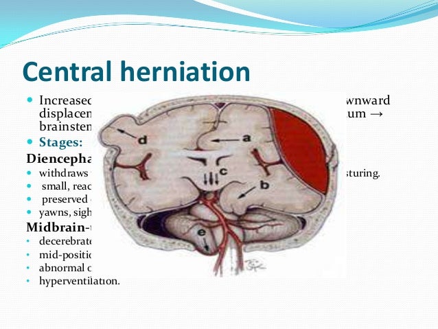
Contralateral Cerebral Peduncle. Herniation of the uncus results in compression of the ipsilateral occulomotor nerve and ipsilateral pupillary dilation [1, 2]; The brain displacement (white arrows) would cause compression of the contralateral cerebral peduncle against the free tentorial edge, as well as the kinking of the contralateral internal carotid artery against rigid structures at the cranial base (black arrows), giving rise to cerebral ischemia of the contralateral hemisphere involving the regions supplied. Mri scans in 20 parkinson�s patients and 20 age and sex matched controls were evaluated in blinded fashion looking for the presence and degree of arterial compression of the cerebral peduncle. Table 3 shows the hemodynamic features of wallerian degeneration.
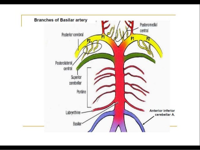
 Posterior circulation stroke Syndromes From slideshare.net
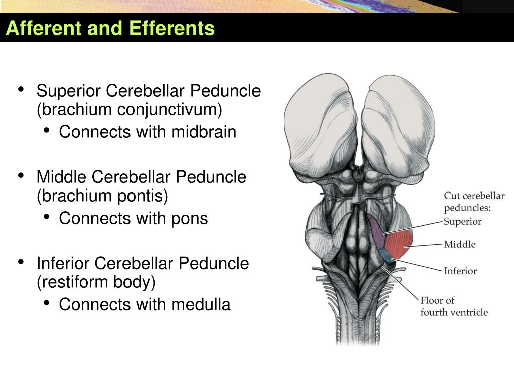
They contain the following afferent fiber tracts: The caudal peduncle contains cerebellar afferent fibers from the lateral cuneate nucleus and dorsal spinocerebellar tract , from vestibular nuclei and nerve , and from the olivary nucleus (climbing fibers). There are six cerebellar peduncles in total, three on each side: A blinded evaluation of mri scans of parkinson�s patients and controls was performed. They returned 18 months later with contralateral peduncle compression. The shift forces the opposing cerebral peduncle against the edge of the tentorium causing contralateral hemiparesis to the peduncle.
The indentation of the tentorium edge causes kernohan�s notch on the opposite cerebral peduncle.
The brain displacement (white arrows) would cause compression of the contralateral cerebral peduncle against the free tentorial edge, as well as the kinking of the contralateral internal carotid artery against rigid structures at the cranial base (black arrows), giving rise to cerebral ischemia of the contralateral hemisphere involving the regions supplied. Early signs of uncal herniation include encroachment on the suprasellar cistern, displacement of the brain stem, enlargement of the ipsilateral crural subarachnoid space, and compression of the contralateral cerebral peduncle. Cerebellar peduncles connect the cerebellum to the brain stem. The only efferent fiber tracts passing through the inferior cerebellar. As the herniating uncus displaces the midbrain laterally, the contralateral cerebral peduncle is compressed against the edge of the tentorium, causing paralysis on the same side as the primary lesion, another false localizing sign. Descending transtentorial herniartion (dth) secondary to a supratentorial mass effect can be detected by ct.
 Source: wjgnet.com
Superior, middle and inferior peduncle afferent fibers of superior cerebellar peduncle. Descending transtentorial herniartion (dth) secondary to a supratentorial mass effect can be detected by ct. Thus hemimegalencephaly or gliomatosis cerebri or widespread cortical dysplasia should be considered. There are six cerebellar peduncles in total, three on each side: This results in weber�s syndrome, which consists of contralateral hemiplegia (including the lower face) due to corticospinal and corticobulbar tract involvement and ipsilateral oculomotor paresis, including parasympathetic cranial nerve iii.
 Source: slideshare.net
A blinded evaluation of mri scans of parkinson�s patients and controls was performed. Caudal cerebellar peduncle (restiform body) connects the cerebellum to the medulla oblongata. Middle cerebellar peduncles connect the cerebellum to the pons and are composed entirely of centripetal fibers. The brain displacement (white arrows) would cause compression of the contralateral cerebral peduncle against the free tentorial edge, as well as the kinking of the contralateral internal carotid artery against rigid structures at the cranial base (black arrows), giving rise to cerebral ischemia of the contralateral hemisphere involving the regions supplied. The middle peduncle is purely afferent.
 Source: what-when-how.com
The following measures were calculated for each patient: They contain the following afferent fiber tracts: The middle peduncle is purely afferent. Table 3 shows the hemodynamic features of wallerian degeneration. The shift forces the opposing cerebral peduncle against the edge of the tentorium causing contralateral hemiparesis to the peduncle.
 Source: doctorlib.info
This is a false localising sign. The inferior cerebellar peduncles are composed of a large restiform body and a small juxtarestiform body. It is major afferent pathway. Herniation of the uncus results in compression of the ipsilateral occulomotor nerve and ipsilateral pupillary dilation [1, 2]; The middle peduncle is purely afferent.
 Source: slideshare.net
They returned 18 months later with contralateral peduncle compression. A blinded evaluation of mri scans of parkinson�s patients and controls was performed. Diffusion tensor imaging (dti) and transcranial magnetic stimulation (tms) could be useful for exploring the state of the corticospinal tract (cst). The middle cerebellar peduncle, or the brachium pontis, enters the cerebellum fairly laterally. Thus hemimegalencephaly or gliomatosis cerebri or widespread cortical dysplasia should be considered.
 Source: slideshare.net
It also contains the bilaterally projecting fibers from the nucleus reticularis tegmenti pontis. Superior, middle and inferior peduncle afferent fibers of superior cerebellar peduncle. Cerebellar peduncles connect the cerebellum to the brain stem. Here we describe a young chinese male patient fulfilling the clinical features of mills’ syndrome, with atrophy of contralateral cerebral peduncle and pontine base without any defined etiology. As the herniating uncus displaces the midbrain laterally, the contralateral cerebral peduncle is compressed against the edge of the tentorium, causing paralysis on the same side as the primary lesion, another false localizing sign.
 Source: slideserve.com
Diffusion tensor imaging (dti) and transcranial magnetic stimulation (tms) could be useful for exploring the state of the corticospinal tract (cst). The middle peduncle is purely afferent. A lesion affecting the cerebral peduncle, especially the medial peduncle, may damage pyramidal fibers and the fascicle of cranial nerve iii. Middle cerebellar peduncles connect the cerebellum to the pons and are composed entirely of centripetal fibers. The inferior cerebellar peduncles are composed of a large restiform body and a small juxtarestiform body.
 Source: thejns.org
Thus hemimegalencephaly or gliomatosis cerebri or widespread cortical dysplasia should be considered. A blinded evaluation of mri scans of parkinson�s patients and controls was performed. They contain the following afferent fiber tracts: There are six cerebellar peduncles in total, three on each side: Thus hemimegalencephaly or gliomatosis cerebri or widespread cortical dysplasia should be considered.
This site is an open community for users to submit their favorite wallpapers on the internet, all images or pictures in this website are for personal wallpaper use only, it is stricly prohibited to use this wallpaper for commercial purposes, if you are the author and find this image is shared without your permission, please kindly raise a DMCA report to Us.
If you find this site convienient, please support us by sharing this posts to your own social media accounts like Facebook, Instagram and so on or you can also save this blog page with the title contralateral cerebral peduncle by using Ctrl + D for devices a laptop with a Windows operating system or Command + D for laptops with an Apple operating system. If you use a smartphone, you can also use the drawer menu of the browser you are using. Whether it’s a Windows, Mac, iOS or Android operating system, you will still be able to bookmark this website.
Category
Related By Category
- Pictures of ethiopian new year
- Snoopy and new baby pics
- Lemon balm metaphysical properties
- Teeth worm
- Nigerian traditional wedding outfits with colour lemon green
- Insetti piccolissimi simili a pidocchi
- Acconciatura capelli corti
- Capellilunghi biondi
- Reign mary
- Melania trump in her wedding gown photos