Cuneiform bone projection
Home » » Cuneiform bone projectionYour Cuneiform bone projection images are ready. Cuneiform bone projection are a topic that is being searched for and liked by netizens now. You can Download the Cuneiform bone projection files here. Find and Download all free photos.
If you’re searching for cuneiform bone projection images information linked to the cuneiform bone projection keyword, you have pay a visit to the ideal site. Our website always provides you with suggestions for viewing the maximum quality video and picture content, please kindly search and locate more enlightening video articles and images that fit your interests.
Cuneiform Bone Projection. The appendicular skeleton is composed of the 126 bones of the appendages and the pectoral and pelvic girdles, which attach the limbs to the axial skeleton. However, joints between the fourth and fifth metatarsals and cuboid bone have greater mobility, which is. A projection or bump with a roughened surface, generally smaller than a tuberosity. Saddle bone deformity or a metatarsal cuneiform exostosis refers to the condition in which a bone on top of the foot is sticking out or protruding.
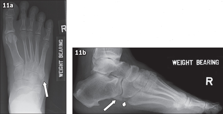
 Common accessory ossicles of the foot imaging features From smj.org.sg
A relatively long, thin projection or bump. The cuneiform bones are the three bones in the foot’s medial side that articulate with the navicular proximally through three separate facets. Another process allows for the attachment of a muscle or ligament. It is grooved inferiorly by the tendon of the flexor hallucis longus m. Medial and lateral borders of the 3 rd (lateral) cuneiform should align with medial and lateral borders of 3 rd metatarsal. The three cuneiform bones are named by location:
The cuneiform bones articulate with the navicular bone proximally and the bases of the metatarsal bones distally:
The cuneiform bones are the three bones in the foot’s medial side that articulate with the navicular proximally through three separate facets. A relatively long, thin projection or bump. The cuneiform bones articulate with the navicular bone proximally and the bases of the metatarsal bones distally: A projection or bump with a roughened surface, generally smaller than a tuberosity. Metatarsal bones these five long bones in the foot incorporate a shaft, a distal head, and a proximal base(27). A projection is an area of a bone that projects above the surface of the bone.
 Source: meded.psu.ac.th
They articulate with the first, second, and third metatarsals, respectively. A relatively long, thin projection or bump. If the case is a fracture without having to prioritize the joint The largest of the cuneiforms. The incidence of this bone is unclear.
 Source: droualb.faculty.mjc.edu
Another process allows for the attachment of a muscle or ligament. A projection that contacts an adjacent bone. Projection clearly demonstrates the scaphoid fracture but the lateral view shows the. The navicular bone articulates with the three cuneiform bones, medial, intermediate, and lateral, to form this joint. The three cuneiform bones are named by location:
 Source: smj.org.sg
A projection is an area of a bone that projects above the surface of the bone. Medial cuneiform (1st) articulates with the 2nd, 3rd, and 4th metatarsal. Medial border of 2 nd metatarsal is aligned with medial border of 2 nd (intermediate) cuneiform. They articulate with the first, second, and third metatarsals, respectively. While it is asymptomatic, it may be a source of discomfort for some people, especially when one feels pain when wearing a shoe.
 Source: slideserve.com
There are 2 varieties of injuries, namely, Less prominent projection located directly inferior to the anterior. It is a shelf of bone that articulates with and supports the talus; A projection that contacts an adjacent bone. Medial border of 2 nd metatarsal is aligned with medial border of 2 nd (intermediate) cuneiform.
This site is an open community for users to do submittion their favorite wallpapers on the internet, all images or pictures in this website are for personal wallpaper use only, it is stricly prohibited to use this wallpaper for commercial purposes, if you are the author and find this image is shared without your permission, please kindly raise a DMCA report to Us.
If you find this site serviceableness, please support us by sharing this posts to your favorite social media accounts like Facebook, Instagram and so on or you can also bookmark this blog page with the title cuneiform bone projection by using Ctrl + D for devices a laptop with a Windows operating system or Command + D for laptops with an Apple operating system. If you use a smartphone, you can also use the drawer menu of the browser you are using. Whether it’s a Windows, Mac, iOS or Android operating system, you will still be able to bookmark this website.
Category
Related By Category
- Pictures of ethiopian new year
- Snoopy and new baby pics
- Lemon balm metaphysical properties
- Teeth worm
- Nigerian traditional wedding outfits with colour lemon green
- Insetti piccolissimi simili a pidocchi
- Acconciatura capelli corti
- Capellilunghi biondi
- Reign mary
- Melania trump in her wedding gown photos